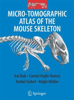
Micro-Tomographic Atlas of the Mouse Skeleton
The Micro-Tomographic Atlas of the Mouse Skeleton provides a unique systematic description of all calcified components of the mouse. It includes about 200 high resolution, two and three dimensional m CT images of the exterior and interiors of all bones and joints. In addition, the spatial relationship of bones within complex skeletal units (e.g., skull, thorax, pelvis, extremities) is also described. The images are accompanied by detailed...
Format:Hardcover
Language:English
ISBN:0387392548
ISBN13:9780387392547
Release Date:September 2007
Publisher:Springer
Length:205 Pages
Weight:2.33 lbs.
Dimensions:0.7" x 8.2" x 10.5"
Customer Reviews
0 rating


